The 12-lead ECG is a fundamental diagnostic tool in cardiology, providing detailed insights into cardiac electrical activity․ Its evolution from early electrocardiography has refined accuracy and clinical utility․ Consisting of 12 electrodes strategically placed on limbs and chest, it captures comprehensive heart activity․ Proper placement ensures precise waveforms, enabling accurate diagnoses of arrhythmias and ischemia․ This guide covers essential aspects of 12-lead ECG placement, including history, components, and clinical applications․
1․1․ History and Evolution of 12-lead ECG
The 12-lead ECG originated from Willem Einthoven’s invention of the first electrocardiograph in 1903, which used three limb leads․ Over the decades, the system evolved to include chest leads, resulting in the 12-lead standard․ This advancement enhanced the ability to detect cardiac abnormalities more accurately․ The modern 12-lead ECG combines limb and chest electrodes to provide a comprehensive view of the heart’s electrical activity․ Its widespread adoption in clinical practice has been driven by its diagnostic precision and non-invasive nature․ Today, it remains a cornerstone in cardiology, aiding in the early detection of conditions like ischemia and arrhythmias․ Standardization and technological improvements continue to refine its utility in patient care․
1․2․ Components of a 12-lead ECG System
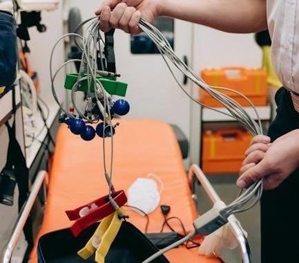
A 12-lead ECG system consists of electrodes, lead wires, and an ECG machine․ The electrodes are placed on the patient’s skin to capture electrical signals from the heart․ These signals are transmitted through insulated wires to the ECG machine, which processes and displays them as waveforms․ The system includes 12 electrodes: 6 chest leads (V1-V6) and 6 limb leads (I, II, III, aVR, aVL, aVF)․ Adhesive electrodes are used for better contact and stability․ The ECG machine amplifies, filters, and records the electrical activity, producing a printout or digital display․ Proper functioning of each component is essential for accurate and reliable readings; Regular maintenance ensures optimal performance and patient safety․
1․3․ Purpose and Scope in Medical Diagnosis
The primary purpose of a 12-lead ECG is to assess the electrical activity of the heart, aiding in the diagnosis of cardiac conditions․ It is non-invasive and provides immediate results, making it a vital tool in emergency and routine care․ The scope extends to identifying arrhythmias, myocardial ischemia, infarction, and hypertrophy․ It also guides pacemaker and implantable cardioverter-defibrillator assessments․ By offering a comprehensive view of heart function, the 12-lead ECG is essential in clinical decision-making, enabling timely interventions․ Its versatility and reliability make it a cornerstone in both acute and chronic cardiac management․ Accurate placement ensures precise diagnostics, emphasizing its critical role in patient care․
Importance of Correct Placement
Correct 12-lead ECG placement is crucial for accurate diagnosis, preventing errors, and ensuring patient safety․ Proper electrode positioning ensures precise waveforms, avoiding misdiagnosis and inappropriate treatment․ Accurate placement is vital․

2․1․ Role in Accurate Diagnosis
Accurate 12-lead ECG placement is pivotal for reliable diagnostic outcomes․ Misplacement can distort waveforms, leading to incorrect interpretations and potential misdiagnoses․ Proper electrode positioning ensures precise capture of P, QRS, and T waves, essential for identifying arrhythmias, ischemia, and other cardiac conditions․ Consistent placement across patients and over time is critical for comparative analysis․ Technicians must adhere to standardized guidelines to minimize variability and maximize diagnostic accuracy․ Errors in placement can obscure crucial electrical activity, underscoring the importance of meticulous technique․ Thus, correct placement directly impacts patient care and treatment decisions․
2․2․ Key Benefits of Proper Placement
Proper 12-lead ECG placement ensures accurate and reliable diagnostic results, enhancing clinical decision-making․ Correct electrode positioning minimizes artifacts and electrical interference, providing clear waveforms for precise interpretation․ It enables early detection of cardiac abnormalities, such as ischemia or arrhythmias, improving patient outcomes․ Consistent placement across patients and over time allows for reliable comparative analysis․ Accurate placement also reduces the need for repeat tests, saving time and resources․ Additionally, it enhances patient safety by preventing misdiagnoses and ensuring appropriate treatment plans․ Proper placement is foundational to high-quality care, making it a critical skill for healthcare professionals․ Its benefits extend to both patients and healthcare systems, promoting efficient and effective cardiac assessment․
2․3․ Limitations and Potential Errors
Despite its diagnostic value, 12-lead ECG placement has limitations and potential errors․ Improper electrode positioning can lead to inaccurate readings, misdiagnoses, or failure to detect critical conditions․ Muscle artifacts, electrical interference, and poor skin preparation are common issues that degrade signal quality․ Obesity, anatomical variations, and movement during the test can also complicate accurate placement․ Additionally, incorrect use of electrodes or adhesives may cause discomfort or skin irritation․ Errors in lead placement, such as reversing limb electrodes, can alter waveform interpretations․ These challenges underscore the importance of proper training and adherence to standardized protocols to minimize errors and ensure reliable results․ Addressing these limitations is crucial for optimizing diagnostic accuracy and patient care․

Standard Placement Guidelines
Standard 12-lead ECG placement involves precise electrode positioning to ensure accurate readings․ Guidelines emphasize proper preparation, electrode selection, and systematic placement to minimize errors and artifacts․ Consistency is key․
3․1․ Preparing the Patient
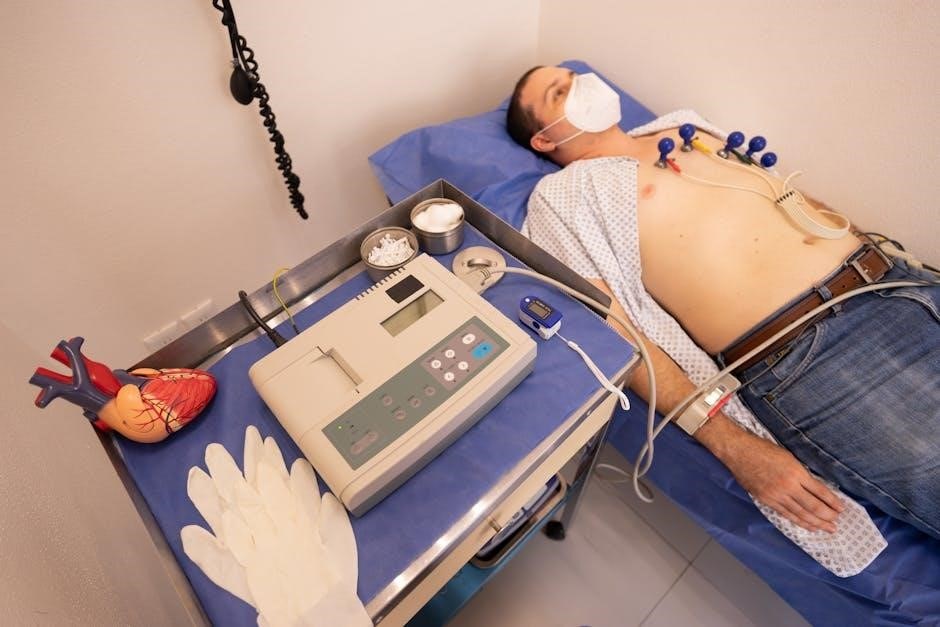
Proper patient preparation is crucial for accurate 12-lead ECG results․ Begin by exposing the chest and all four limbs to ensure electrode placement accessibility․ Remove any jewelry or clothing that may interfere with electrode adhesion․ Clean and dry the skin to enhance electrode contact, avoiding oils or lotions․ Position the patient in a comfortable, supine position with arms relaxed at their sides and legs uncrossed to minimize movement artifacts․ Ensure the room is quiet and the patient is calm to reduce electrical interference․ Verify the patient’s identity and medical history before proceeding․ These steps ensure optimal conditions for precise ECG recording and interpretation․
3․2․ Types of Electrodes and Adhesives
12-lead ECG electrodes are typically made of silver/silver chloride, known for their excellent conductivity and low interference․ Most electrodes are precoated with a conductive gel to ensure proper adhesion and signal quality․ There are also foil electrodes for specific situations, such as pediatric or sensitive skin patients, which can be non-allergenic․ Additionally, carbon-based electrodes are available for prolonged monitoring․ Adhesives play a critical role in securing electrodes; hypoallergenic options are recommended for sensitive skin․ Proper storage and handling of electrodes are essential to maintain their effectiveness․ Always follow manufacturer guidelines for electrode placement and adhesive use to ensure accurate ECG recordings and patient comfort․
3․3․ Step-by-Step Placement Process
Begin by preparing the patient, ensuring skin is clean and dry for optimal electrode adhesion․ Apply limb electrodes first, positioning them on the upper arms and thighs, avoiding bony areas․ Chest electrodes are placed next, starting with V1 at the fourth intercostal space to the right of the sternum․ V2 mirrors V1 on the left, while V3-V6 are placed across the chest in a horizontal line․ Ensure each electrode is securely attached with conductive gel and adhesive․ Double-check all placements for consistency and accuracy, adjusting as needed for patient anatomy․ Finally, connect the leads to the ECG machine and verify signal quality before recording․ Proper placement ensures reliable results․

Anatomical Considerations
Accurate 12-lead ECG placement requires identifying correct anatomical landmarks, such as intercostal spaces and limb positions․ Gender differences, like chest lead placement in females, must be considered․ Proper body positioning ensures electrode alignment for precise recordings․ Consistency is key to avoid signal distortion and ensure reliable diagnostic results․
4․1; Identifying Correct Landmarks
Identifying correct anatomical landmarks is crucial for accurate 12-lead ECG placement․ The fourth intercostal space at the midclavicular line is the reference point for V1 and V2․ For chest leads (V1-V6), proper alignment with the intercostal spaces ensures precise recording of cardiac activity․ Limb leads require placement on the upper arms and thighs, avoiding bony structures․ Palpation of the clavicle and sternum helps locate the correct intercostal spaces․ For female patients, chest leads (V3-V6) should be placed under the breast tissue to ensure proper contact and signal quality․ Consistent landmark identification minimizes variability and ensures reliable ECG waveforms for accurate diagnosis․ Proper technique is essential for obtaining high-quality recordings, especially in diverse patient anatomies․
4․2․ Gender Differences in Placement
Gender differences play a significant role in 12-lead ECG placement, particularly for chest leads․ In female patients, leads V3 to V6 should be placed under the breast tissue to ensure proper contact and avoid interference․ This adjustment accounts for anatomical variations and prevents signal distortion․ For males, these leads are typically placed directly on the chest wall․ Additionally, the clavicle and sternum landmarks remain consistent across genders, but technicians must be mindful of breast tissue placement in females to maintain accuracy․ Proper gender-specific adjustments ensure high-quality ECG recordings and accurate diagnoses․ Awareness of these differences is critical for technicians to avoid errors and ensure reliable results․
4․3․ Body Positioning for Accuracy
Proper body positioning is crucial for accurate 12-lead ECG recordings․ Patients should lie supine with their arms relaxed at their sides and legs slightly abducted to minimize muscle tension․ The chest and limbs must be fully exposed to facilitate electrode placement․ For optimal signal quality, the patient should remain still and avoid muscle contractions, as movement artifacts can distort waveforms․ Breathing should be calm and regular, avoiding deep breaths that may cause baseline shifts․ In some cases, elevating the upper body slightly may improve comfort without compromising accuracy․ Ensuring the patient is relaxed and properly positioned minimizes artifacts and ensures reliable ECG results, which are essential for precise cardiac evaluations․

Limb Leads Placement
Limb leads are placed on the upper arms and thighs, avoiding bone․ Electrodes must be uniform, ensuring accurate electrical activity capture․ Consistency prevents diagnostic errors․
5․1․ Upper Limb Electrode Placement
Upper limb electrodes are placed on the arms, typically 2-3 cm above the wrist crease on the lateral aspect․ Avoid placing electrodes directly over bone or tendons․ For male patients, ensure the electrode is not placed under the sleeve or cuff․ Clean and prepare the skin to ensure proper adhesion and signal quality․ The right arm (RA) electrode is placed on the right wrist, while the left arm (LA) electrode is placed on the left wrist․ Ensure symmetry between the arms for accurate readings․ The reference electrode (RL) is often placed on the chest or upper limb․ Proper placement ensures clear signal capture and accurate ECG interpretation․
5․2․ Lower Limb Electrode Placement
Lower limb electrodes are placed on the anterior aspect of the thighs, typically 2-3 cm above the knee․ Ensure electrodes are not placed directly over bone or joints․ The right leg (RL) and left leg (LL) electrodes should be symmetrically positioned to maintain consistency․ Clean and prepare the skin to ensure proper adhesion and signal quality․ Avoid placing electrodes too close to the knee or ankle to prevent movement artifacts․ Proper placement ensures clear signal capture and accurate ECG interpretation․ This method aligns with standard guidelines for 12-lead ECG placement, ensuring reliable diagnostic results․ Symmetry and correct positioning are crucial for minimizing interference and ensuring accurate readings․
5․3․ Common Mistakes to Avoid
One of the most frequent errors in 12-lead ECG placement is inconsistent electrode positioning, which can lead to inaccurate readings․ Mislabeling electrodes or swapping their positions is another common mistake, causing confusion and misdiagnosis․ Improper skin preparation, such as failing to clean or dry the skin, can result in poor adhesion and signal interference․ Placing electrodes too close to joints or bones should be avoided, as this can introduce artifacts․ Additionally, neglecting to ensure symmetry in limb lead placement can distort waveform accuracy․ Awareness of these pitfalls is crucial for obtaining reliable ECG results․ Healthcare professionals must adhere to standardized guidelines to minimize errors and ensure diagnostic precision․
Chest Leads Placement
Chest leads placement involves strategically positioning electrodes V1 to V6 across the torso to capture heart activity from multiple angles․ Proper landmarks and consistency are critical for accurate readings, ensuring electrodes are placed correctly to avoid artifacts and misdiagnosis․
6․1․ V1 to V6 Lead Placement
V1 to V6 leads are placed across the chest to capture the heart’s electrical activity from a horizontal plane․ V1 is positioned at the fourth intercostal space to the right of the sternum, while V2 is placed mirroring V1 on the left side․ V3 is located midway between V2 and V4, ensuring proper alignment․ V4 is placed at the fifth intercostal space in the midclavicular line, with V5 and V6 positioned laterally, maintaining consistency in spacing․ Correct placement avoids muscle and bone interference, ensuring clear waveforms for accurate diagnoses․ Special considerations, such as placing V3-V6 under the breast in female patients, are crucial for optimal results․

6․2․ Ensuring Consistency
Consistency in chest lead placement is critical for accurate ECG interpretation․ V1 to V6 leads must align with anatomical landmarks, such as the midclavicular line, to ensure uniformity․ Proper spacing between leads prevents overlap and ensures clear waveforms․ For female patients, V3 to V6 leads should be placed under the breast tissue to avoid interference․ Using standardized reference points, such as the fourth and fifth intercostal spaces, helps maintain consistency across patients․ Regular training and adherence to guidelines minimize variability․ Consistency ensures reliable comparisons in serial ECGs, which is vital for detecting subtle changes in cardiac activity․ Proper documentation of lead placement further enhances diagnostic accuracy and clinical decision-making․
6․3․ Special Adjustments Needed
Chest lead placement may require special adjustments for accurate readings․ For female patients, V3 to V6 leads should be placed under the breast tissue to ensure proper contact․ In obese patients, leads may need to be positioned more laterally or higher to avoid excess tissue interference․ Pediatric patients often require adjusted placements due to smaller chest sizes․ Additionally, leads should avoid bony prominences to prevent interference․ Proper electrode adhesion is crucial, especially in sweaty or hairy areas․ These adjustments ensure reliable ECG waveforms and accurate diagnoses․ Regular training and adherence to guidelines help in making these modifications effectively, ensuring consistency and reliability in ECG readings across diverse patient populations․
Special Considerations
Special considerations in 12-lead ECG placement involve pediatric, neonatal, and obese patients, requiring adjusted electrode sizing and positioning․ Movement artifacts must be managed for accurate readings․
7․1․ Pediatric and Neonatal Patients
Pediatric and neonatal patients require special attention in 12-lead ECG placement due to their smaller body size and delicate skin․ Electrodes should be scaled down to fit appropriately, ensuring comfort and proper adhesion․ For neonates, electrodes are often placed on the upper arms and thighs instead of wrists and ankles․ Chest leads, particularly V1 to V6, must be carefully positioned to avoid interference from breathing movements․ Consistency in placement is crucial to maintain accurate readings․ Additionally, minimizing movement during the procedure is essential to reduce artifacts․ Proper training is necessary to handle these patients effectively, ensuring high-quality ECG recordings for accurate diagnosis․
7․2․ Placement in Obese Patients
ECG placement in obese patients requires careful consideration to ensure accurate readings․ Excess body fat can interfere with electrode adhesion and signal quality․ Larger or specialized electrodes may be necessary to accommodate thicker skin and ensure proper contact․ Landmarks for electrode placement should be identified through palpation rather than visualization, as anatomical references may be obscured․ Limb electrodes should be placed on the upper arms and thighs to avoid excess fat layers․ Chest electrodes, particularly V1 to V6, may need to be adjusted slightly higher to account for breast tissue․ Consistency in placement is crucial, and additional electrodes or alternative adhesives may be required to maintain contact․ Proper training is essential to handle these challenges effectively․
7․3․ Managing Movement Artifacts
Movement artifacts are common challenges during ECG recordings, especially in restless or uncooperative patients․ To minimize these interferences, ensure the patient remains still and relaxed during the procedure․ Use high-quality electrodes with strong adhesion to maintain proper contact․ For patients with excessive movement, consider using electrodes with built-in motion reduction features or securing them with additional adhesive strips․ Instruct the patient to avoid talking, coughing, or deep breathing during the recording․ If artifacts persist, reassess electrode placement and ensure all leads are firmly attached․ In pediatric or neonatal cases, swaddling or using non-invasive restraints may help․ Consistency and patience are key to obtaining clear, artifact-free tracings․

Troubleshooting Common Issues
Identify and correct electrode misplacement, loose connections, or poor adhesion․ Ensure proper patient preparation and minimize movement artifacts․ Verify lead placement and connection accuracy for clear tracings․
8․1․ Identifying and Correcting Errors
Identifying errors in 12-lead ECG placement is crucial for accurate diagnostics․ Common issues include electrode misplacement, loose connections, or improper adhesion․ Muscle artifacts or electrical interference may distort waveforms․ To correct errors, recheck electrode positions, ensuring adherence to standard guidelines․ Adjust leads to avoid skin folds or bony prominences․ Verify the ECG machine’s calibration and patient preparation․ Documenting discrepancies and recalibrating as needed ensures reliable results․ Proper troubleshooting minimizes repeat tests, saving time and improving patient care․ Regular training and competency assessments help technicians master error identification and correction, enhancing overall ECG accuracy and clinical decision-making․
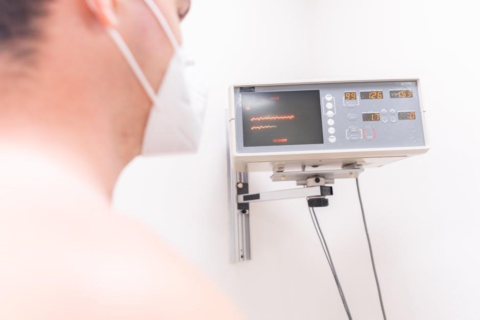
8․2․ Interpreting Artifacts
Artifacts in 12-lead ECGs are non-physiological signals that distort waveform accuracy․ Common causes include muscle tremors, electrical interference, and poor electrode adhesion․ Identifying artifacts involves analyzing irregular patterns, such as baseline wander or sudden spikes․ Correcting these issues often requires adjusting electrode placement, ensuring proper skin preparation, or using noise-reduction filters․ Regular equipment calibration also minimizes artifacts․ Accurate interpretation is vital to avoid misdiagnosis․ Technicians should document and address artifacts promptly to ensure reliable ECG results․ By understanding the sources and solutions, healthcare professionals can enhance the clarity and diagnostic value of 12-lead ECG recordings, improving patient care outcomes․

Training and Competency
Effective training ensures proper 12-lead ECG placement, enhancing diagnostic accuracy․ Programs include hands-on practice, simulations, and competency assessments to confirm proficiency in electrode placement and artifact reduction․
9․1․ Training Programs
Structured training programs are essential for mastering 12-lead ECG placement․ These programs typically include hands-on sessions, simulations, and interactive modules to ensure proficiency․ Participants learn electrode placement, patient preparation, and how to minimize artifacts․ The curriculum often covers anatomical landmarks, gender-specific adjustments, and special considerations for pediatric and obese patients․ Practical exercises allow trainees to apply theoretical knowledge, while feedback from instructors helps refine techniques․ Many programs incorporate case studies to demonstrate real-world applications, enhancing diagnostic accuracy․ Successful completion of these programs equips healthcare professionals with the skills to perform ECGs confidently and accurately, ensuring reliable diagnostic results․
9․2․ Competency Assessment
Competency assessment ensures healthcare professionals master 12-lead ECG placement, verifying their ability to perform accurate and reliable recordings․ This process involves rigorous evaluation of electrode placement skills, adherence to anatomical landmarks, and understanding of patient preparation․ Practical exams, where technicians demonstrate electrode placement on mannequins or actual patients, are common assessment methods․ Standardized checklists are used to evaluate accuracy, ensuring all steps are correctly followed․ Additionally, written tests may assess knowledge of ECG principles and troubleshooting․ Feedback from instructors helps refine techniques, while periodic re-assessment maintains high standards․ Competency assessment is crucial for ensuring accurate diagnoses and patient safety, making it a cornerstone of ECG training programs․
Digital Resources and References
Reputable sources like the American Heart Association offer comprehensive PDF guides and online modules for mastering 12-lead ECG placement․ Utilize Boolean operators in searches for precise results․
10․1․ Recommended PDF Guides
Several authoritative PDF guides are available for mastering 12-lead ECG placement․ The American Heart Association (AHA) offers detailed manuals with diagrams and step-by-step instructions․ Philips Healthcare and GE HealthCare provide comprehensive guides emphasizing correct electrode positioning․ These resources include visual aids and troubleshooting tips․ Additionally, educational institutions and medical training websites publish free PDF guides tailored for students and professionals․ When searching, use specific terms like “12-lead ECG placement PDF” or “ECG electrode placement guide” to find reliable materials․ These guides are essential for ensuring accurate placements and interpreting results effectively․ They are widely accessible online, supporting both clinical practice and educational needs․
10․2․ Online Training Modules
Online training modules offer interactive and accessible learning for 12-lead ECG placement․ Platforms like Coursera, Udemy, and medical education websites provide structured courses with videos, quizzes, and downloadable resources․ These modules cover theoretical knowledge and practical skills, ensuring proficiency in electrode placement and ECG interpretation․ Many programs are designed for healthcare professionals, students, and EMTs, catering to varying skill levels․ They often include case studies and real-world scenarios to enhance learning․ Searching for “ECG placement online course” or “12-lead ECG training modules” yields numerous options․ These resources are ideal for continuous education and competency development, ensuring accurate and reliable ECG recordings in clinical settings․
The proper placement of a 12-lead ECG is vital for accurate diagnosis and effective patient care․ This guide has covered essential aspects, from historical evolution to modern techniques, emphasizing the importance of correct electrode positioning․ By adhering to standardized guidelines and understanding anatomical considerations, healthcare professionals can minimize errors and ensure reliable results․ Continuous training and the use of digital resources are key to maintaining competency․ Proper 12-lead ECG placement not only enhances diagnostic accuracy but also improves patient outcomes․ This comprehensive overview serves as a valuable reference for anyone seeking to master the skills required for precise and effective ECG recording․